European School for Advanced Veterinary Studies
Individual Courses and Comprehensive Study Program
Ultrasonography – Introduction to Ultrasonography
The objectives of this course are to provide participants with a basic knowledge of ultrasound image formation and current technology, and to perform routine abdominal ultrasonographic examination in dogs and cats.
Main topics
- Basic principle of ultrasonographic image formation
- Ultrasonographic unit: transducer and setting
- Semiology and artifacts
- General principles of abdominal US examination
- Liver, spleen and lymph nodes
- Female genital tract
- Urinary bladder, male genital tract
- GIT, pancreas and adrenal glands
- Introduction to US guided biopsies and aspiration
- Doppler principles
- Anatomy of the abdominal vessels
Diagnostic Ultrasound I
This course is meant for veterinarians who already have some practical experience in ultrasound. The aim is to sharpen the participant’s perception and to discuss ultrasound images of various pathological conditions.
Topics:
- Instrumentation / techniques / artifacts
- Approach and interpretation
- Abdomen
- Endosonography
- Biopsy / interventional sonography
- Diagnostic value of US compared to other methods
- General including abdominal wall
- Liver
- GI: including oesophagus and pancreas
- Urinary tract
- Genital tract
- Vascular including Doppler of arteries and veins
- Lymph nodes
- Adrenal system
Diagnostic Ultrasound II
The aim of this advanced course is to further sharpen the participant’s perception and to discuss ultrasound images of various pathological conditions.
Topics:
- Advanced diagnostic ultrasound technologies and different approaches
- Interventional ultrasound
- Doppler basics
- Portosystemic shunts
- Non-cardiac thoracic ultrasound
- Urogenital tract
- Endocrine ultrasound
- Other parenchymal organs
- Musculoskeletal ultrasound
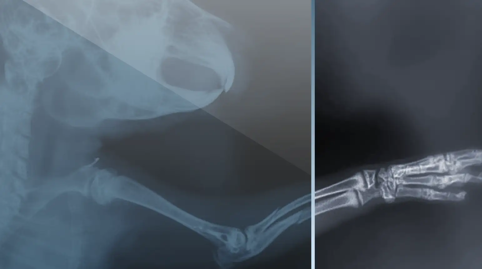
Radiology I
This course is designed for veterinarians with limited experience in diagnostic radiology of companion animals. The objective is to provide participants with a logical approach to basic radiographic image interpretation, including detection of pertinent radiographic signs, and generation of a prioritized differential diagnosis.
Topics:
- Thorax – Introduction to interpretation of radiographic images
- Thoracic radiology
- Pulmonary patterns
- Abdomen – Radiographic contrast media and contrast procedures
- Appendicular skeleton – Aggressive versus non-aggressive bone lesion
- Axial skeleton – Myelography
Radiology II
This advanced course is designed for veterinarians who wish to broaden their knowledge in diagnostic radiology of companion animals.
Topics:
- Thorax (advanced) – Thoracic radiology
- Pulmonary patterns
- Abdomen (advanced) – Radiographic contrast media and contrast procedures
- Appendicular skeleton (advanced) – Aggressive versus non-aggressive bone lesion
- Axial skeleton (advanced) – Myelography
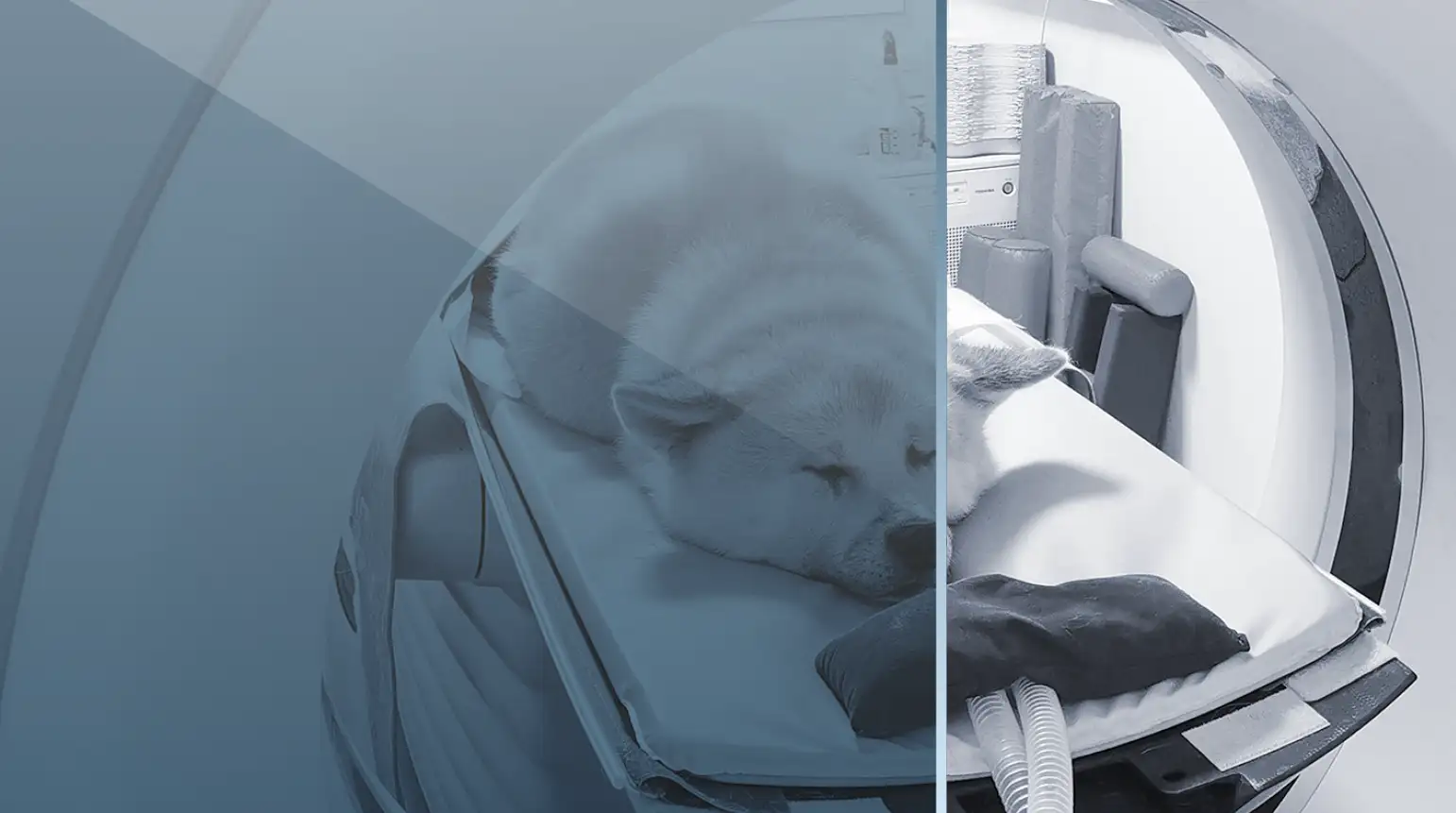
Small Animal Computed Tomography
This course is designed for veterinarians who have access to computed tomography in their daily practice and wish to be able to prescribe, acquire and interpret routine CT studies. The objectives are to provide participants with a basic understanding of the technology, knowledge of common CT acquisition techniques, proficiency in image viewing and manipulation, and a logical approach to CT image interpretation.
Topics:
- Basic principles, instrumentation and acquisition principles
- Image reconstruction, quality, and artifacts
- DICOM, PACS, viewing stations and image display
- Orthopaedics
- Neuroimaging – Spine and CNS
- Head CT, thoracic CT
- CT angiogram and vascular anatomy – Abdomen
Neuropathology and MRI Interpretation
This interactive course welcomes neurologists, pathologists, radiologists, diagnostic imagers and other MRI users as well as trainees in these disciplines. The principal learning objectives are to acquire a basic understanding of diagnostic neuropathology and to apply this knowledge to the interpretation of MR images.
Topics:
- Practical gross neuroanatomy in MRI and brain sections
- Recognizing gross lesion patterns
- Recognizing microscopic lesion patterns
- Principles of classification and interpretation of lesions
- Principles of MRI reading